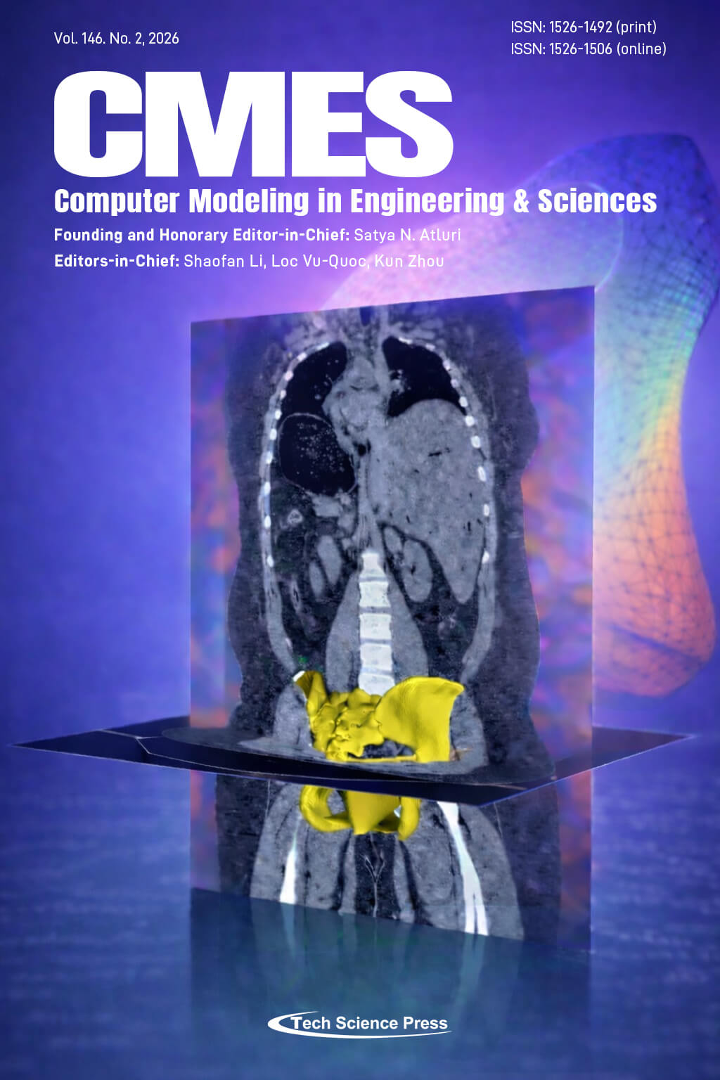
The image illustrates an integrated imaging–modeling framework for the human pelvic region, combining medical imaging, anatomical detail, and computational statistical shape analysis. The transition from CT-based anatomical imaging to a reconstructed pelvic geometry highlights the data-driven personalization of pelvic bones and floor muscles. The layered visualization depicts shape prediction and non-rigid mesh alignment. Overall, the image conveys the fusion of anatomy derived from medical imaging and statistical shape modeling, which facilitates the understanding of pelvic structure and function.
The background was created by AI and contains no human likenesses, copyrighted elements, or misleading representations.
View this paper

 Submit a Paper
 Propose a Special lssue